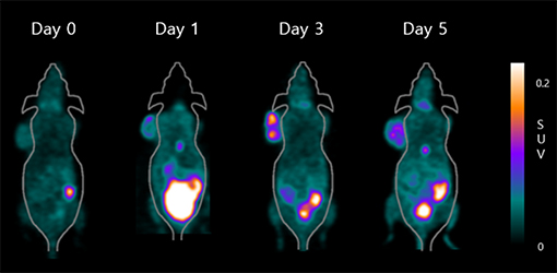
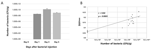
Tumor-targeting bacteria have been actively investigated as a new therapeutic tool for solid tumors. However, in vivo imaging of tumor-targeting bacteria has not been fully established. [18F]fluorodeoxysorbitol (FDS) positron emission tomography (PET) is known to be capable of imaging Gram-negative Enterobacteriaceae infection. We validated the use of [18F]FDS PET for visualization of the colonization and proliferation of tumor-targeting Escherichia coli (E. coli ) in mouse tumor models.

18F-FDS PET imaging of tumor-bearing mice treated with E. coli.

(A) Viable bacterial counts in harvested tumors.
(B) Correlation between SUVmax and the number of viable bacteria in tumors.
[18F]FDS PET showed the distribution of tumor-targeting E. coli, even in the deep portions of tumors, and provided semiquantitative data on bacterial location without sacrificing the mice. Therefore, [18F]FDS PET has potential as a promising in vivo imaging technique for visualizing the therapeutic process when bacterial cancer therapy (BCT) is performed with Enterobacteriaceae such as E. coli.
It has been known that roughly 1,500,000 people die from invasive fungal infection every year and, as the number of patients with HIV/AIDS and those who receive chemotherapy, modern immunosuppressive therapy, and invasive medical interventions are growing, the incidence of invasive fungal infection is also constantly increasing. Radiolabeled sorbitol, 2-deoxy-2-[18F]fluorosorbitol ([18F]FDS), initially evaluated for the diagnosis of glioma in mice, has been studied for imaging pathogenic E. coli. It is intriguing that sorbitol is also used as a sole carbon source supporting high fungal growth and sporulation. In conclusion, [18F]FDS showed specific targeting and prolonged retention in A. fumigatus infection with low background activity. It suggests that this radiotracer could be used as a novel PET molecular imaging agent to obtain outstanding image quality in the diagnosis of A. fumigatus infection.